Electron microscopy and spectroscopy are great tools for peering into matter on the molecular scale. But they’re not terribly effective if that matter happens to be biological.
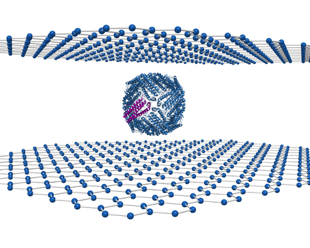
Researchers at the University of Illinois in Chicago (UIC) may have changed that with a novel use of graphene in which a biomolecule is sandwiched between two sheets of graphene, making it possible to create much higher resolution images than in previous methods.
“We found a way to encapsulate a liquid sample in two very thin layers of graphene — sheets of carbon that are only one atom thick,” said Canhui Wang, UIC graduate student in physics and first author of the study, in a press release.
In the research, which was published in the journal Advanced Materials (“High-Resolution Electron Microscopy and Spectroscopy of Ferritin in Biocompatible Graphene Liquid Cells and Graphene Sandwiches”), the molecule ferritin was imaged. Ferritin is an iron-storage protein, and itself has been proposed for developing magnetic nanoparticles to enable forward osmosis for water purification.
Prior to this research, if you wanted to image a biomolecule like ferritin with an electron microscope, you would need to use a container known as a “liquid stage” that is wedged between two thick windows of silicon nitrate to protect the sample from the vacuum.
Robert Klie, the senior investigator on the study, likened the difference between the liquid stage approach and the graphene sandwich as “…the difference between looking through Saran Wrap and thick crystal.”
The better resolution produced by the graphene sandwich is not just because of graphene’s superior transparency. The graphene sandwich also provides better protection from the electron beam that is fired at a sample during microscopy. Wang says that some have calculated that to visualize a sample requires 10 times the amount of radiation one would be exposed to from a 10-megaton hydrogen bomb when standing just 30 meters away.
To mitigate the deleterious effect of the electron beam, most electron microscopes use a low-energy beam that results in a fuzzy picture that has to be corrected by imaging algorithms. Because of graphene’s high thermal and electrical conductivity the material removes both the heat and electrons generated by the beam as it passes through the sample. So higher energy electron beams can be used on the sample, resulting in higher resolution images.
In their experiments with the ferritin, the researchers were able to image for the first time iron oxide in the core of ferritin changing its electric charge, leading to the release of iron. The imaging of this process could lead to a better understanding of some human disorders.
“Defects in ferritin are associated with many diseases and disorders, but it has not been well understood how a dysfunctional ferritin works towards triggering life-threatening diseases in the brain and other parts of the human body,” said Tolou Shokuhfar, assistant professor of mechanical engineering-engineering mechanics at Michigan Technological University and adjunct professor of physics at UIC, in a press release.


