Researchers have used one of the brightest X-ray sources on the planet to map the 3-D structure of an important cellular gatekeeper known as a G protein-coupled receptor, or GPCR, in a more natural state than possible before. The new technique is a major advance in exploring GPCRs, a vast, hard-to-study family of proteins that plays a key role in human health and is targeted by an estimated 40 percent of modern medicines.
The research, performed at the Linac Coherent Light Source (LCLS) X-ray laser at the Department of Energy's (DOE's) SLAC National Accelerator Laboratory, is also a leap forward for structural biology experiments at LCLS, which has opened up many new avenues for exploring the molecular world since its launch in 2009.
"For the first time we have a room-temperature, high-resolution structure of one of the most difficult to study but medically important families of membrane proteins," said Vadim Cherezov, a pioneer in GPCR research at The Scripps Research Institute who led the experiment. "And we have validated this new method so that it can be confidently used for solving new structures."
In the experiment, published in the Dec. 20 issue of Science, researchers examined the human serotonin receptor, which plays a role in learning, mood and sleep and is the target of drugs that combat obesity, depression and migraines. The scientists prepared crystallized samples of the receptor in a fatty gel that mimics its environment in the cell. With a newly designed injection system, they streamed the gel into the path of the LCLS X-ray pulses, which hit the crystals and produced patterns used to reconstruct a high-resolution, 3-D model of the receptor.

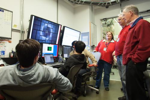
A team led by Vadim Cherezov of The Scripps Research Institute views an X-ray diffraction pattern, produced when SLAC's Linac Coherent Light Source X-ray laser struck a crystallized sample of the human serotonin receptor. The team collected and analyzed more than 100,000 of these patterns to reproduce a 3-D structure of the receptor. Credit: Fabricio Sousa/SLAC
The method eliminates one of the biggest hurdles in the study of GPCRs: They are notoriously difficult to crystallize in the large sizes needed for conventional X-ray studies at synchrotrons. Because LCLS is millions of times brighter than the most powerful synchrotrons and produces ultrafast snapshots, it allows researchers to use tiny crystals and collect data in the instant before any damage sets in. As a bonus, the samples do not have to be frozen to protect them from X-ray damage, and can be examined in a more natural state.

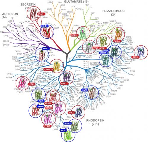
This diagram, a "family tree" for G protein-coupled receptors, highlights the receptors (circled) whose structures have been solved, among hundreds of others whose structures have not yet been determined. Credit: GPCR Network:
"This is one of the niches that LCLS is perfect for," said SLAC Staff Scientist Sébastien Boutet, a co-author of the report. "With really challenging proteins like this you often need years to develop crystals that are large enough to study at synchrotron X-ray facilities."


