Neurotransmitters play a powerful role in managing the sympathetic and parasympathetic nervous system in many animals, controlling mood, energy levels, aggression, social bonding, and sexual behavior. For example, many antidepressant drugs modulate the uptake of serotonin, a neurotransmitter associated with mood.

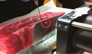
Nanoparticles with a "reporter molecule" are injected into a slab of lamb. These molecules can be detected through the use of Raman spectroscopy. The red circle indicates the location where a laser light was focused onto the lamb bone. (Courtesy of ACS).
Nanoparticles with a "reporter molecule" are injected into a slab of lamb. These molecules can be detected through the use of Raman spectroscopy. The red circle indicates the location where a laser light was focused onto the lamb bone. (Courtesy of ACS).
While neurotransmitters play a pivotal role in the nervous systems of many organisms, monitoring and determining levels of neurotransmitters in the brain can be challenging. To measure the level of neurotransmitters through the use of existing techniques, researchers have to drill a hole through a patient's skull. However, some new technologies promise a less-invasive way to monitor and study neurotransmitters.
According to a new study published by the American Chemical Society, Raman spectroscopy can be used to monitor levels of neurotransmitters in the brain. With this form of spectroscopy, researchers can look for neurotransmitters and can detect chemical signatures that are associated with a variety of materials. For example, the technology could be used to detect chemical signatures linked to explosives. In another study, researchers used Raman spectroscopy to monitor a living rat's glucose levels through its skin (in a non-invasive way).
To detect molecules through calcified bone, researchers used both Raman spectroscopy with spatially offset and surface-enhanced techniques. Both of these techniques involve the excitation of samples through the application of a high-power laser. Once samples reach a predefined excitation state, researchers monitor the samples for these Raman signals.
With the spatially offset method, researchers are able to successfully detect a signal from molecules that are embedded up to 20 millimeters inside a solid sample. Signals on these embedded molecules can be isolated by measuring Raman signals at an offset from the penetration point of the laser light. This offset ensures that photon scattering doesn't impact the signal to noise ratio of the Raman signal too significantly.
Surface-enhanced Raman spectroscopy comprises the use of gold nanoparticles increase signals that are produced by molecules that are bound to a surface.
In the study, researchers used a slab of lamb shoulder with a bone that had a varying thickness of three to eight millimeters. In comparison, the average human skull has a thickness that varies from three to 14 millimeters. Once the meat slab was loaded into the spectroscopy system, 90 trillion gold nanoparticles were injected behind the bone. These particles were saturated with a compound that exhibited a vigorous Raman signature. When researchers shined a 785-nm laser on the bone they were immediately able to detect chemical signatures for the reporter molecule.
That said, the technologies can't locate nanoparticles inside tissue; current Raman spectroscopy limitations prevent deep tissue penetration.


